Is this inspection method applicable to all 3D printing materials, including plastics and ceramics?
X-ray and CT inspection can cover metals, plastics, and ceramics, but no single setup applies to every material, size, wall thickness, or defect type. Density, attenuation, thickness, geometry, contrast, voxel sampling, surface condition, and access determine detectability. Select 2D radiography or 3D CT from the material and acceptance question; the method does not automatically qualify every part or service case. For an RFQ, provide the alloy or polymer, ceramic grade, process, dimensions, channels, lot, final condition, inspection criterion, and report needs.
Universal Principles with Material-Specific Optimization
The physics is material-dependent. Metals usually need higher energy and artifact control, plastics may offer lower attenuation but weaker contrast, and ceramics can combine high density with brittle or complex geometry. Compare attenuation, thickness, feature access, and expected defect contrast before choosing the scanner, detector, exposure, and reconstruction method.
Inspection Principle: X-ray attenuation depends on material density and atomic number. This means we adjust voltage, current, and filtering to achieve optimal contrast for each material family, from low-density polymers to high-density metals and ceramics.
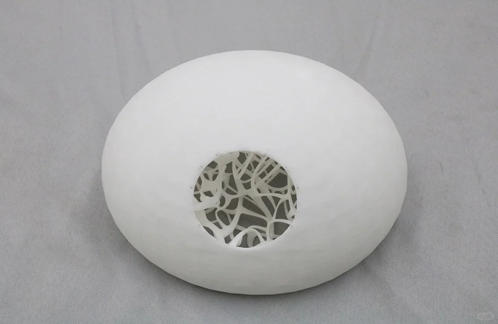
Plastic and Polymer Material Inspection
Plastic parts can be screened for voids, inclusions, trapped material, and dimensional or channel conditions when the contrast supports detection. Record the polymer, additive process, moisture or conditioning state, wall thickness, and final condition. A low-density image can still miss a low-contrast feature, so verify the method with a reference and acceptance rule.
Technical Adaptation:
Higher kVp Settings: Enhanced penetration for carbon-filled composites and engineering plastics
Specialized Detectors: High contrast resolution for identifying voids in Nylon (PA) and Polycarbonate (PC)
Defect Sensitivity: Capable of detecting pores as small as 10 microns in Polyether Ether Ketone (PEEK)
Reportable Metrics for Plastics:
Internal void percentage and distribution
Fiber orientation analysis in composite materials
Layer adhesion quality from Material Extrusion processes
Wall thickness variations in hollow structures
Ceramic Material Inspection Capabilities
Ceramic inspection may require higher energy, careful fixturing, and a reference that reflects the material and geometry. Review cracks, pores, inclusions, density changes, and channels with the appropriate CT or radiography route. The scan is evidence for the defined inspection scope; it does not replace mechanical, phase, or functional testing when those are required.
Technical Adaptation:
High Energy CT Systems: For penetrating dense Zirconia (ZrO₂) and Alumina (Al₂O₃)
Phase-Contrast Imaging: Enhanced detection of micro-cracks in Silicon Carbide (SiC)
Subsurface Defect Mapping: Critical for Ceramic components in medical and aerospace applications
Reportable Metrics for Ceramics:
Sintering density uniformity
Micro-crack density and propagation
Granular porosity from binder burnout processes
Density gradient analysis
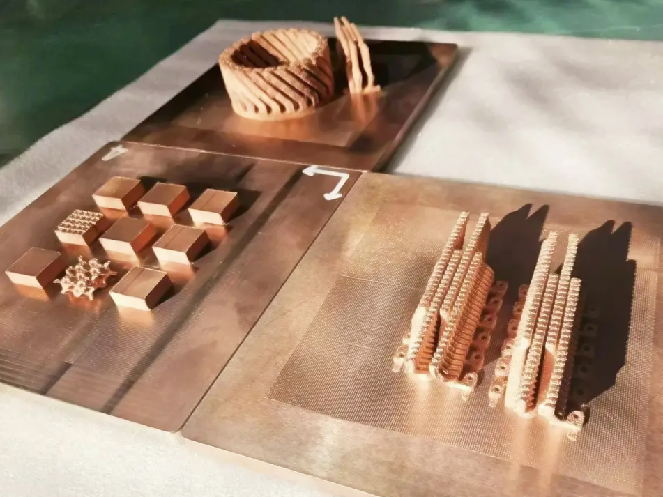
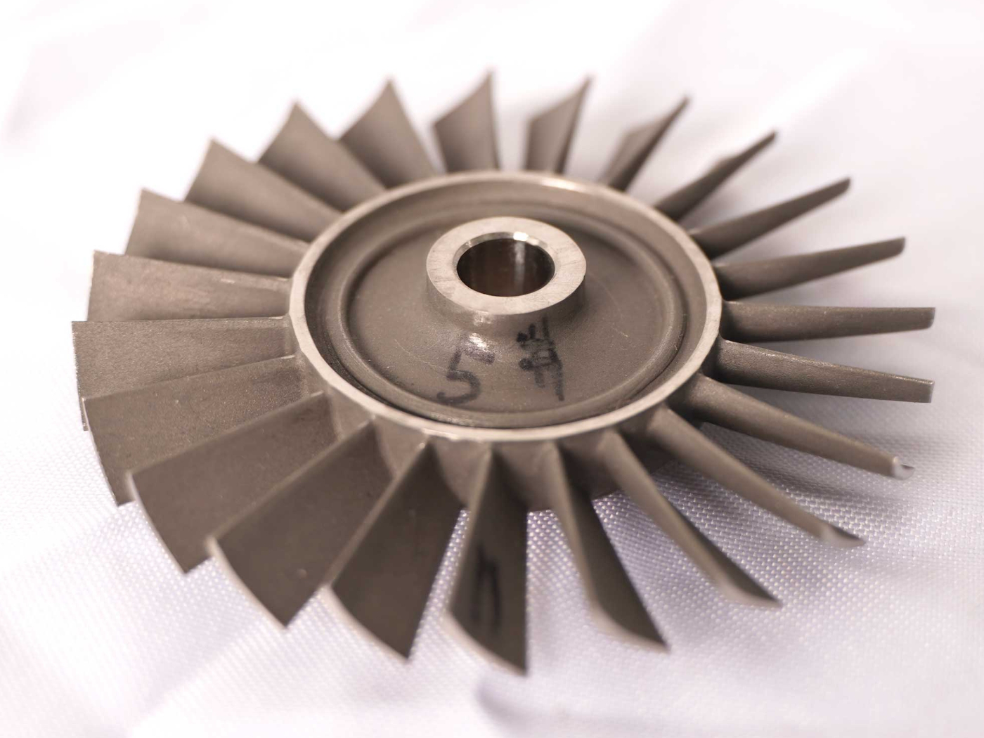
Metallic Material Excellence
Metal AM parts often need attention to powder-related porosity, lack-of-fusion, inclusions, cracks, support interfaces, and internal channels. Choose the scan direction and resolution from the alloy, wall thickness, geometry, orientation, and service load. Keep as-built, heat-treated, HIP-treated, and machined states separate, then inspect the final condition against the drawing.
Technical Adaptation:
Multi-energy CT for alloy composition verification
High-resolution micro-CT for critical Aerospace and Aviation components
Correlation with Heat Treatment effectiveness
Material-Specific Highlights:
Titanium Alloy: Alpha-case formation detection
Aluminum Alloys: Hydrogen porosity quantification
Stainless Steel: Inclusion content analysis
Superalloy: Hot isostatic pressing effectiveness verification
Advanced Resin and Photopolymer Analysis
For Vat Photopolymerization components, we've developed specialized low-energy protocols.
Technical Adaptation:
Low-energy X-ray settings to prevent material damage
Staining techniques for enhanced contrast in clear Resins
UV degradation assessment in aged components
Industry-Specific Validation Requirements
The suitable method also depends on the use case. A prototype may need screening, while an aerospace, medical, automotive, or energy part may require a qualified reference, quantitative report, repeatability evidence, and an approval review. State the acceptance criterion and verification method before treating the result as release evidence.
Medical Device Manufacturing:
100% inspection of Medical and Healthcare implants from CP-Ti (Grade 1-4)
Pore size limits according to ASTM F2884 for laser-sintered components
Automotive and Aerospace:
Process qualification for Automotive safety components
Lot-to-lot consistency monitoring for production parts
Inspection scope should match the material and geometry. For parts made with Plastic 3D Printing, Ceramic 3D Printing, or metal additive manufacturing, provide the CAD or drawing revision, size and wall range, internal features, quantity, final state, critical defects, resolution target, acceptance standard, and schedule. We can then select the route, identify limitations, and record the disposition.
Material and Geometry Limits
X-ray methods can be used for metals, plastics, and ceramics, but attenuation and contrast change with density, composition, thickness, porosity, and geometry. A setup that works for a small polymer housing may not resolve an indication through a thick nickel alloy wall. Ceramic parts may also require care because cracks, pores, and density gradients can have similar image contrast.
Provide material, dimensions, wall thickness, internal channels, expected defect type, critical zones, and acceptance threshold before selecting the scan. Confirm whether the result is qualitative screening, quantitative defect sizing, dimensional metrology, or a combination. The report should separate detected indications from areas that could not be evaluated reliably.